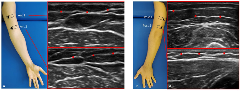
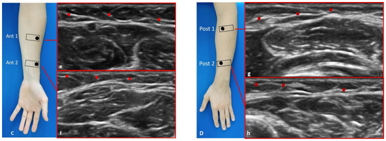
Figure 1.
Ultrasound (US) images of the superficial fascia of: the anterior region of the arm (A) and of the forearm (C); the posterior region of the arm (B) and of the forearm (D). Anterior regions (A,C) at levels Ant 1 (a,e) and Ant 2 (b,f). Posterior regions (B,D) at levels Post 1 (c,g) and Post 2 (d,h). Probe: black rectangle. Red dashes: superficial fascia. Reprinted with permission from [15].


