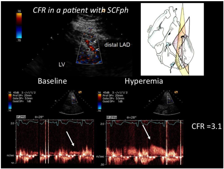
Figure 2.
CFR in the distal LAD assessed by E-Doppler TTE in a patient with SCFph. At the top, color flow in the distal LAD (in red); on the right, a cartoon of the tomographic plane orientation to obtain the LAD insonification. At the bottom, pulsed Doppler spectral tracing of the blood flow velocity in the distal LAD at baseline (left) and at maximal Adenosine-induced hyperemia (right); note the prevalent diastolic BF velocity, a peculiarity of coronary flow. The CFR (peak hyperemic diastolic velocity/peak resting diastolic velocity) is above 3, indicating no significant stenosis and a fairly normal endothelium-independent microcirculatory function.

