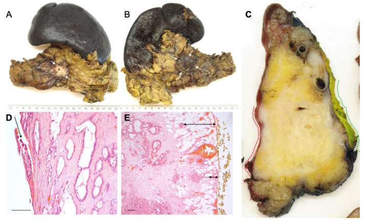
Figure 3.
Involvement of the anterior pancreatic surface and Gerota’s fascia by this large tumour in the pancreatic body is suspected macroscopically, both on external inspection ((A) anterior surface, arrows, and (B) posterior surface with Gerota’s fascia, arrows) and on a sagittal specimen slice ((C) anterior surface and Gerota’s fascia, red and green dotted lines, respectively). Histology confirms tumour breaching of the anterior surface ((D) arrow) and infiltration within 1 mm of the posterior surface of Gerota’s fascia ((E) arrows). Magnification: 400× (D), 200× (E). Scale bar (D,E): 200 microns.

