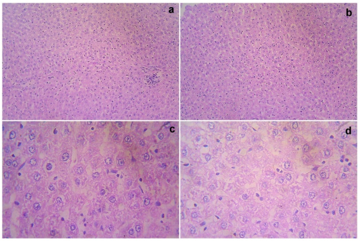
Figure 2.
Gross histopathological examination of the liver following the infusion of mesenchymal stem cells obtained from decidua basalis in Wistar albino rats to assess the impact of safety, efficacy, and acute toxicity. Representative images of the liver section stand for (a) 10× showing central vein and sinusoidal dilatation, (b) 10× showing lobular architecture, (c) 40× showing focal necrosis, and (d) 40× showing cytoplasmic vacuolation and necrosis, respectively.

