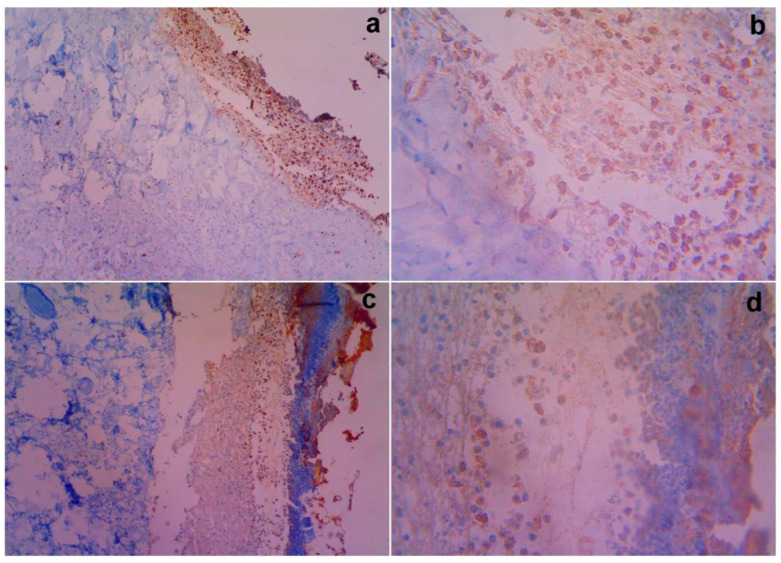
Figure 6.
Gross histopathological examination of the different post excision for COX-2 nuclear and cytoplasmic immunoreactive positivity with treated rats on day 14. Representative images of the COX-2 nuclear and cytoplasmic section stand for (a) 10× showing magnification of the control showing 3(+) immunoreactivity compared with Group II (c) showing 2(+) immunoreactivity for MSCs, (b) 40× showing magnification of 3(+) immunoreactivity by the vehicle control, and (d) 40× showing magnification of 2(+) immunoreactivity by the test article, respectively.

