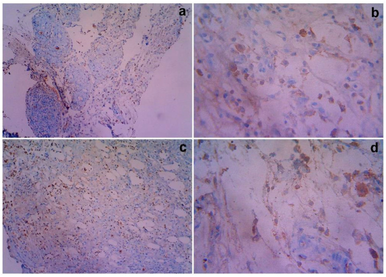
Figure 7.
Gross histopathological examination of the different post excisions for iNOS nuclear and cytoplasmic immuno reactive positivity with treated rats on day 14. Representative images of the iNOS nuclear and cytoplasmic section stand for (a) 10× showing iNOS for the untreated control cytoplasmic positivity (1+) and immunoreactivity (1+), (b). 40× shows granulation tissue shows 1+ respectively. iNOS for MSCs treated group shows Cytoplasmic positivity (2+) and Immunoreactive (2+): (c). 10× shows immuno reactive 2+, (d). 40× shows immuno reactive 2+, respectively.

