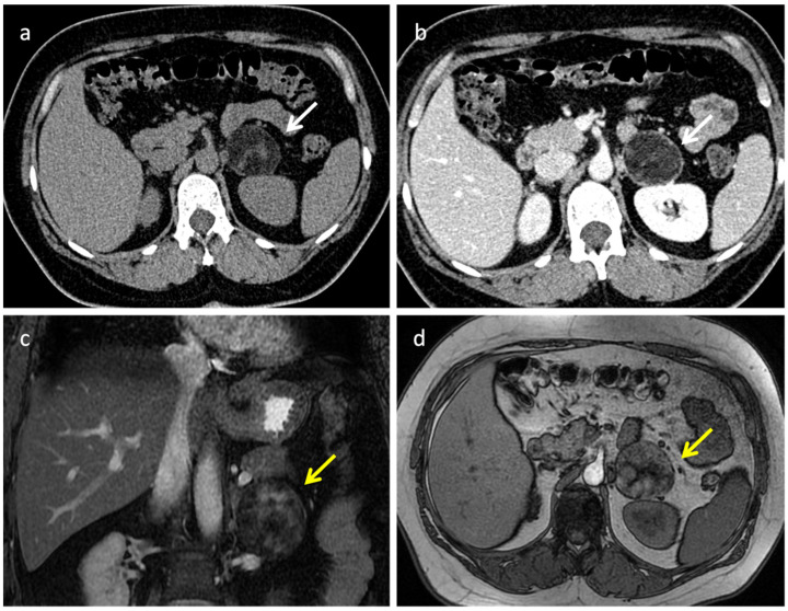
Figure 2.
Adrenal myelolipoma in a 42-year-old woman. CT demonstrates a large mass within the left adrenal gland (white arrow in (a,b) and yellow arrows in (c,d)), characterized by well-defined borders and internal inhomogeneity. Axial unenhanced CT (a) shows attenuation values <−30 HU due to the presence of gross fat. On portal venous phase (b), the mass shows subtle enhancement due to its poor vascularization. Coronal T2 fat sat sequence (c) and axial T1-weighted out-phase image (d) demonstrate extracellular fat.

