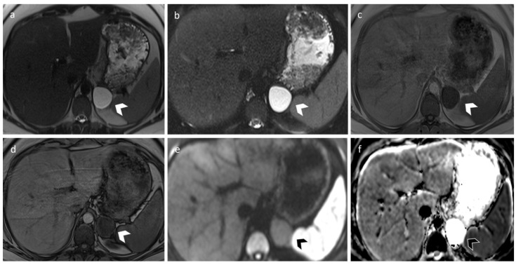
Figure 4.
A 33-year-old female with an incidental left adrenal cyst. MR revealed a 4 cm nodular lesion in the left adrenal gland characterized by thin walls and high and homogeneous signal in T2 (a) and T2 fat-sat sequences (b). T1 signal (c,d) is in accordance with simple fluid content, and DWI/ADC sequence (e,f) does not show signs of hypercellularity.

