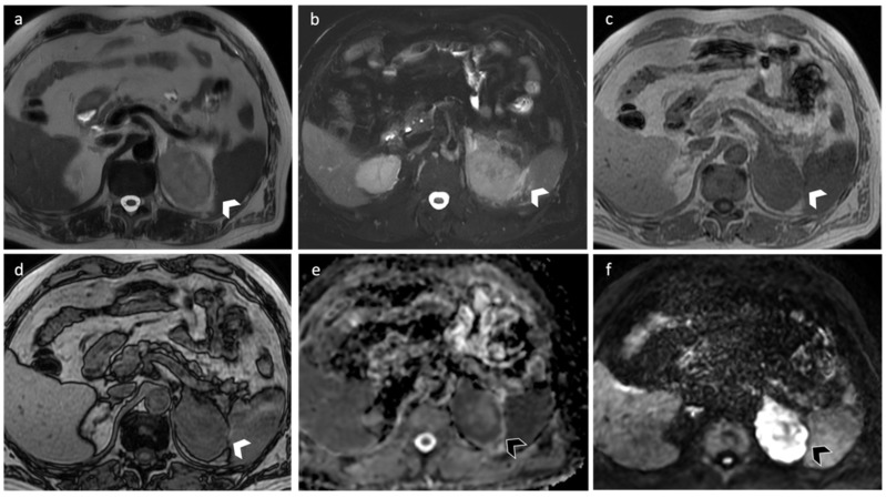
Figure 8.
A 67-year-old man with lung cancer. MRI shows a 7 cm nodular lesion on the left adrenal gland characterized by intermediate signal intensity on axial T2-weighted images (a) and perilesional edema (b). The lesion shows intermediate T1-intensity (c) with no signal loss in out-of-phase sequences (d), due to the absence of intralesional fat. DWI (e) and ADC map (f) demonstrated hypercellularity.

