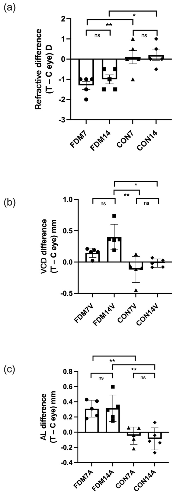
Figure 1.
Differences in ocular refraction (a), vitreous chamber depth (b), and AL (c) between the treated eye and contralateral control eye (left and right eyes in the CON7 and CON14 groups) in the FDM and control groups. The three parameters indicated significant myopia in the FDM eyes compared with the CON7 and CON14 eyes. Values are shown as the mean ± standard error. * p < 0.05, ** p < 0.01. C, contralateral control eye; ns, not significant; T, treated eye; VCD, vitreous chamber depth.

