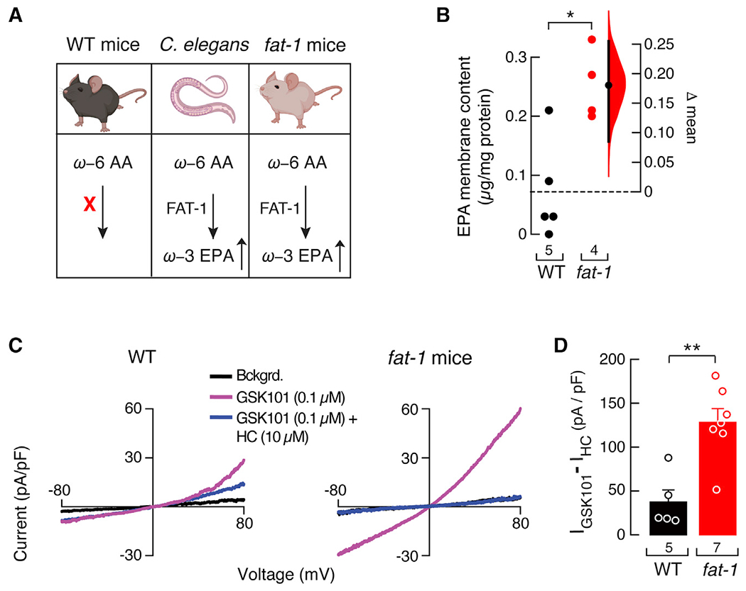
Figure 2. Isolated endothelial cells from fat-1 mice have increased TRPV4 activity.

(A) The C. elegans fatty acid desaturase FAT-1 enzyme introduces a double bond in ω-6 arachidonic acid to synthesize ω-3 EPA in worms and transgenic fat-1 mice, but not in WT mice. Mice and C. elegans cartoons were created with BioRender.com.
(B) Gardner-Altman estimation plot showing the mean difference in EPA-membrane content of whole mesenteric arteries of WT and fat-1 mice, as determined by LC-MS. The raw data are plotted on the left axis. The mean difference, on the right, is depicted as a dot; the 95% confidence interval is indicated by the ends of the vertical error bars. Mann-Whitney rank test for two independent groups.
(C) Representative current-voltage relationships determined by whole-cell patch-clamp recordings of WT and fat-1 cultured isolated mesenteric endothelial cells challenged with GSK101 (0.1 μM) and GSK101 (0.1 μM) + HC (10 μM). Bckgrd. indicates background currents.
(D) Bar graph displaying TRPV4 currents ((IGSK101 – IHC) pA/pF) obtained by whole-cell patch-clamp recordings (+80 mV) of cultured isolated mesenteric endothelial cells of WT and fat-1 mice. Bars are mean ± SEM. Two-tailed unpaired t test. Asterisks indicate values significantly different from WT (**p < 0.01 and *p < 0.05). n is indicated in each panel.
