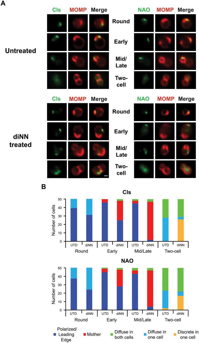
Fig 4. Effect of the CL-targeting antibiotic 3’,6-dinonylneamine (diNN) on the localization of Cls_6xH and aPLs at different stages of the division process.
HeLa cells were infected with the Cls_6xH transformant, and at 22hpi, infected cells were lysed and Cls_6xH expression was induced by incubating the cells in the lysate for 1.5hrs. in axenic media containing 10nM aTc in the presence or absence of 5 μM diNN. Cells were then fixed and the distribution of MOMP, Cls_6xH (labeled Cls), or NAO in chlamydial cells was assessed. (A) Representative images of the indicated markers in Untreated or diNN treated bacteria at the indicated stages of cell division. Images were acquired with a Zeiss AxioImager2 microscope equipped with a 100x oil immersion PlanApochromat lens and are representative of two independent experiments. Scalebar = 2 μm (B) 50 individual cells from the indicated stages of division from untreated (UTD) and diNN-treated cells were assessed for their Cls_6xH and NAO localization profiles. Localization profiles were categorized into polarize/leading edge of daughter cell, mother, diffuse in one cell, diffuse in both cells, or discrete in one cell. The differences in localization of Cls_6xH and NAO between treatment conditions at each stage of division were statistically analyzed using a chi-squared test. This revealed that the changes induced by diNN treatment were statistically significant (p<0.001) for each division intermediate compared to untreated (UTD) controls. There was no statistical difference between Cls_6xH and NAO localization profiles for any condition tested.

