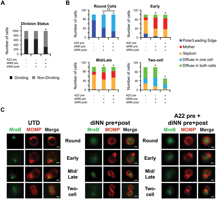
Fig 7. Effect of the CL-targeting antibiotic 3’,6-dinonylneamine (diNN) and the MreB-targeting antibiotic A22 on localization of MreB_6xH.
HeLa cells were infected with the MreB_6xH transformant, and, at 22hpi, infected cells were lysed and chlamydial cells in the lysate were induced by incubating the cells in axenic media containing 10nM aTc. In some instances, cells were preincubated with 5μM diNN or 5μM diNN + 75 μM A22 for 30 minutes and then induced with aTc in the presence of 5μM diNN alone. Following induction, the localization of MreB_6xH at various stages of division was assessed by staining cells with MOMP and 6xHis antibodies. (A) The total number of dividing versus non-dividing cells from the indicated culture conditions was quantified from 1000 total bacteria. * = p<0.0001 compared to the untreated (UTD) control as measured by chi-squared test. (B) The localization of MreB_6xH was assessed in individual cells from each stage of division from untreated cultures or cultures treated as indicated in the figure. Localization profiles were categorized into leading edge of the budding daughter cell/polar, diffuse in mother cell, diffuse in one cell, diffuse in both cells, or septum. The differences in localization of MreB_6xH between treatment conditions at each stage of division were statistically analyzed using a chi-squared test to reveal that the changes resulting from drug treatments were statistically significant when compared to UTD (* = p<0.0001). Pretreatment of cells with diNN and A22 resulted in a statistically significant difference in MreB localization in pre-division intermediates (round cells) when compared to pretreatment with diNN alone (** = p<0.02). For (A) and (B), data were pooled from two independent experiments. (C) Representative images illustrating the distribution of MreB_6xH (labeled MreB) at different stages of division in untreated cells or in cells treated with drugs as indicated in the figure. Scalebar = 2 μm.

