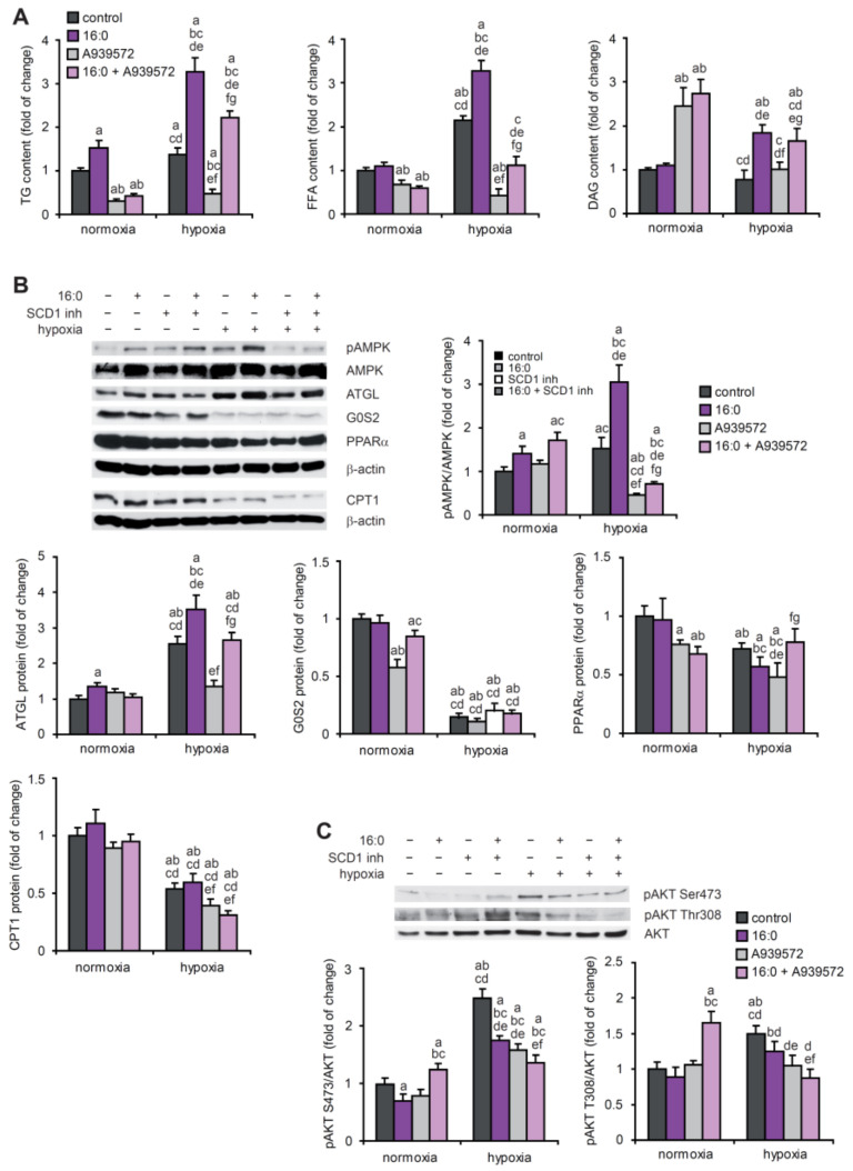
Figure 6.
The effect of SCD1 inhibition and 16:0 treatment on lipid and energy metabolism in hypoxic HL-1 cardiomyocytes. (A) HL-1 cells were treated with 2 µM A939572 (SCD1 inhibitor) and/or 100 µM 16:0 and subjected to hypoxia for 18 h. Neutral lipids were extracted from the cells and separated using the thin layer chromatography method. The content of TG, FFA, and DAG was determined by densitometry. Protein levels of (B) pAMPK, AMPK, ATGL, G0S2, PPARα, and CPT1 and (C) pAKT[Ser473], pAKT[Ser308], and AKT were determined by Western blot. The data are representative of n = 3 independent experiments. The data are expressed as the mean ± SD. a—p < 0.05, vs. control/normoxia; b—p < 0.05, vs. 16:0/normoxia; c—p < 0.05, vs. A939572/normoxia; d—p < 0.05, vs. 16:0 + A939572/normoxia; e—p < 0.05, vs. control/hypoxia; f—p < 0.05, vs. 16:0/hypoxia; g—p < 0.05, vs. A939572/hypoxia (ANOVA).

