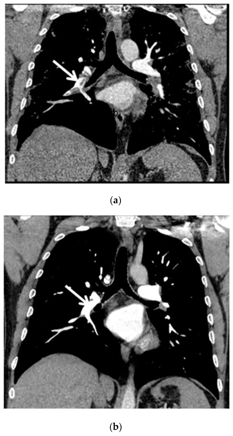
Figure 2.
(a) Chest CT scan (mediastinal window, coronal view) shows pulmonary embolism that affects the right pulmonary artery, lobar arteries of the right lower and upper lobes and interlobar pulmonary artery (white arrow); (b) Chest CT scan after six months of follow-up shows complete resolution of thrombosis (white arrow).

