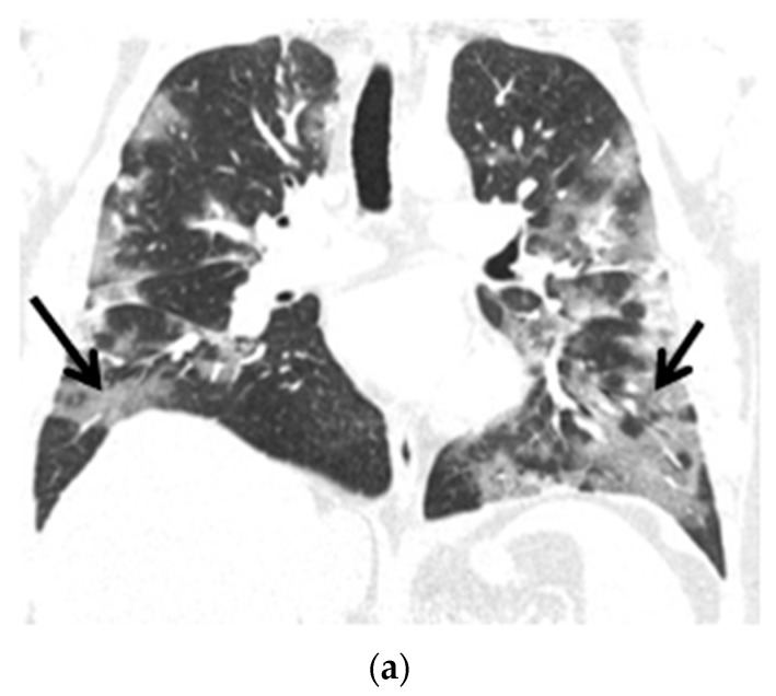
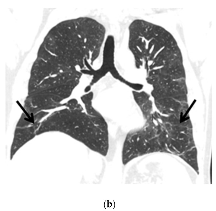
Figure 3.
(a) Chest CT scans (lung window, coronal view) show patchy ground-glass opacities in accordance with COVID-19 dominant in the peripheral zones of the lower lungs (black arrows); (b) Chest CT scans (lung window, coronal view) after six months of follow-up show resolution of lung lesions (black arrows).


