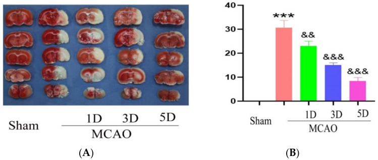
Figure 1.
The cerebral infarction volume were detected with TTC staining method following middle cerebral artery occlusion and treadmill exercise (A) TTC staining method was used to detect brain sections of mouse with middle cerebral artery occlusion and treadmill exercise 1D, 3D and 5D; White is the cerebral infarction area, and red is normal area. (B) Statistical analysis of cerebral infarct volume in (A). (*** p < 0.001 vs. Sham; && p < 0.01, &&& p < 0.001 vs. MCAO) n = 5.

