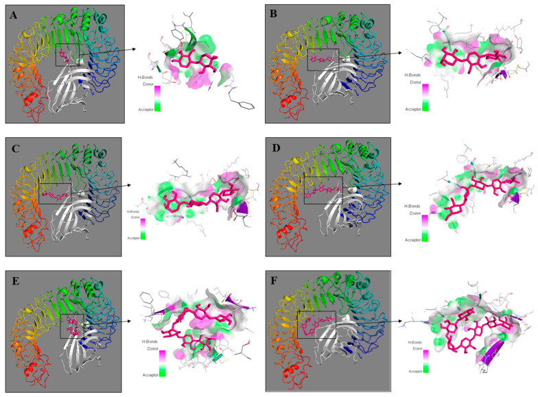
Figure 6.
Three-dimensional structures of molecular docking studies representing binding affinity of xylooligosaccharides—xylobiose (A), xylotriose (B), xylotetraose (C), xylopentaose (D), xylohexaose (E), and xyloheptaose (F) (shown in pink) and TLR4 (chain A) and myeloid differentiation factor-2 (MD-2) (shown in rainbow and white, respectively). Protein–ligand complexes were visualized by Discovery Studio 2016 V16.1.0.

