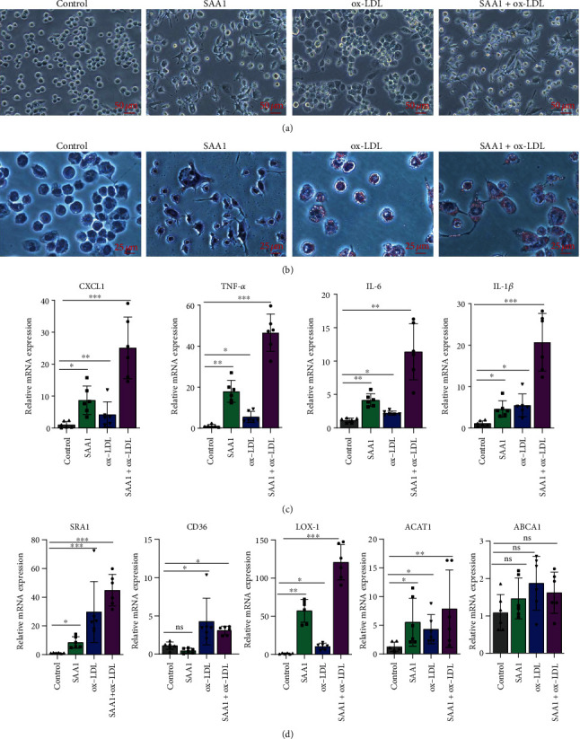
Figure 2.

Effects of SAA1 on activation of macrophages and foam-cell formation. The macrophages derived from THP-1 cells induced by PMA were treated with SAA1, ox-LDL, or SAA1+ox-LDL, with the untreated macrophages as the control. (a) The morphology of macrophages was observed under microscopy. (b) Lipid deposition in cells was assessed by Oil Red O staining. (c) The expression of CXCL1, TNF-α, IL-6, and IL-1β was detected by real-time PCR. (d) The expression of SRA1, CD36, LOX-1, ACAT1, and ABCA1 was detected by real-time PCR. Scale bars, 25 μm and 50 μm. Data represent the mean ± SEM; n = 6 (ns: not significant; ∗P < 0.05, ∗∗P < 0.01, and ∗∗∗P < 0.001).
