Figure 5.

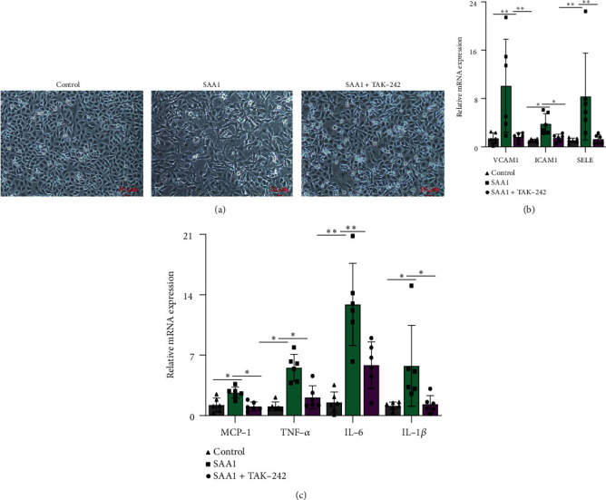
The role of TLR4 in SAA1-induced endothelial inflammatory phenotype. HUVECs were stimulated with SAA1 or SAA1+TAK-242, respectively. (a) Morphological changes in cells were observed by microscopy. (b) The expression of adhesion molecules (VCAM1, ICAM1, and SELE) was detected by real-time PCR. (c) Real-time PCR was used to detect the expression of proinflammatory molecules (MCP-1, TNF-α, IL-6, and IL-1β). Scale bar, 10 μm. Data represent the mean ± SEM; n = 6 (∗P < 0.05 and ∗∗P < 0.01).
