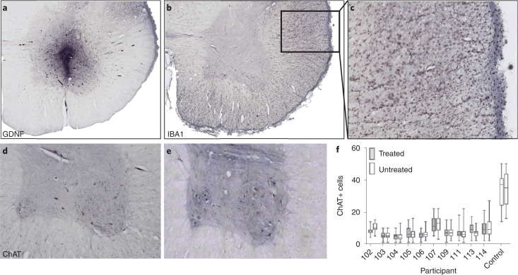
Fig. 4. Motor neurons were similar between treated and untreated sides.
a,b, Immunohistochemistry showed graft survival with a high level of GDNF (a), yet very low levels of IBA1 staining (b). c, Descending motor neuron tracts showed an inflammatory response with hypertrophic microglia and IBA1 staining. d,e, Immunohistochemistry showed that ChAT-positive motor neurons were (d) lost in the ALS lumbar spinal cord and (e) preserved in the control spinal cord. f, The treated and untreated spinal cord sides showed similar numbers of ChAT-positive motor neurons across participants; motor neurons were significantly lower in participant 102 (p = 0.0001) and significantly higher (p = 0.0013) in 113 on the treated compared to untreated side. Box plots depict median and 25th to 75th percentiles, with min and max whiskers. Sample size n = 47 (on average) independent participant spinal cord sections, n = 25 independent control spinal cord sections. Magnification, ×2.5 (a,b); ×20 (c); ×5 (d,e).

