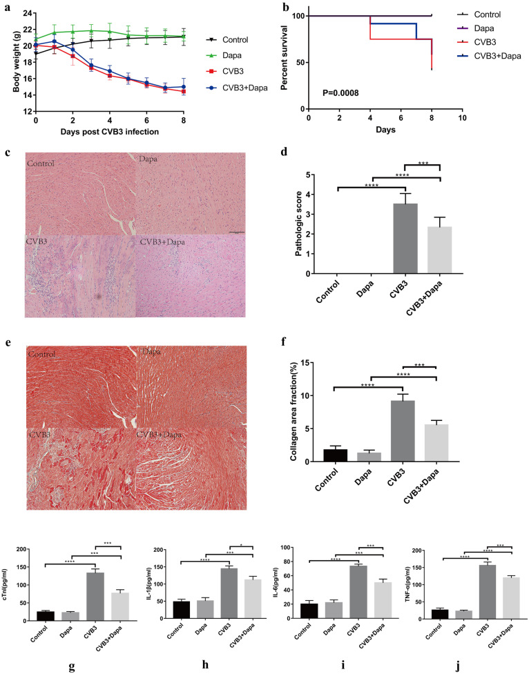
Fig. 1.
Dapagliflozin treatment ameliorates myocarditis. Viral myocarditis model was created in BALB/C mice as described in Methods. Monitor the a body weight change and b survival rate of mice from day 0 to day 8. Survival proportions at day 8 were 100% for the control group and Dapa group. c Hematoxylin–eosin staining to observe the inflammatory response to myocarditis. Red-stained area shows myocardial tissue, blue staining shows inflammatory cell infiltration (magnification: × 200, scale bar: 100 µm). d The severity of myocarditis was scored using a standard 0–4 grading scale. e Masson staining to observe the inflammatory response to myocarditis. Myocardial cells were stained red and collagenous fibers were stained blue (magnification: × 200, scale bar: 100 µm). f Collagen area fraction (collagen area/field area × 100%) was calculated by the Image-Pro Plus analysis system. g Serum myocardial injury markers cardiac troponin I (CTnI) were measured by ELISA. h, i, j Serum inflammation markers IL-1β, IL-6, and TNF-α were measured by ELISA. Data are shown as mean ± SD. (control, normal mice; Dapa, normal mice treated with dapagliflozin; CVB3, CVB3-infected mice treated with 60% propylene glycol; CVB3 + Dapa, CVB3-infected mice treated with dapagliflozin. *P < 0.05, **P < 0.01, ***P < 0.001, ****P < 0.0001).

