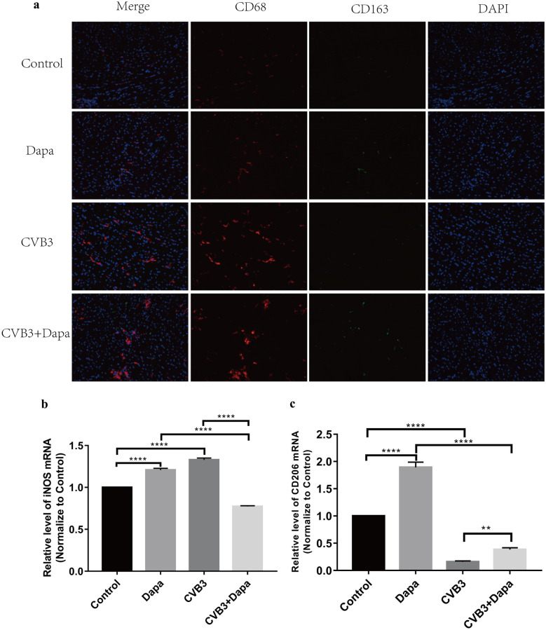
Fig. 2.
Dapagliflozin decreased the percentage of M1 and increased the percentage of M2 in myocardium at day8 after CVB3 infection. a Immunofluorescent double staining of myocardial tissue with antibodies to CD68 and CD163. Red indicates CD68 (one marker for M1), green CD163 (one marker for M2), and blue DAPI-stained cellular nuclei. b, c Quantitative reverse transcription polymerase chain reaction (qRT-PCR) was used to detect iNOS and CD206 mRNA levels. Data were expressed as mean ± SD (control, normal mice; Dapa, normal mice treated with dapagliflozin; CVB3, CVB3-infected mice treated with 60% propylene glycol; CVB3 + Dapa, CVB3-infected mice treated with dapagliflozin. *P < 0.05, **P < 0.01, ***P < 0.001, ****P < 0.0001).

