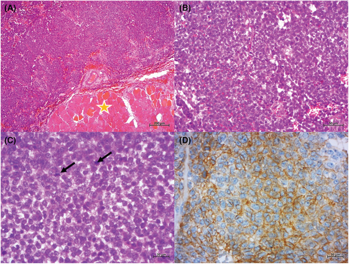
FIGURE 1.

(A) Biopsy specimen showing a highly cellular tumor infiltrating the skeletal muscle (asterisk) (H&Ex50); (B) the tumor is composed of uniform small round cells with hyperchromatic nuclei and indistinct cytoplasmic borders (H&Ex200); (C) note the presence of mitosis (arrows) and the absence of osteoid production (H&Ex400); (D) tumor cells show a diffuse membranous staining for CD99 (x400)
