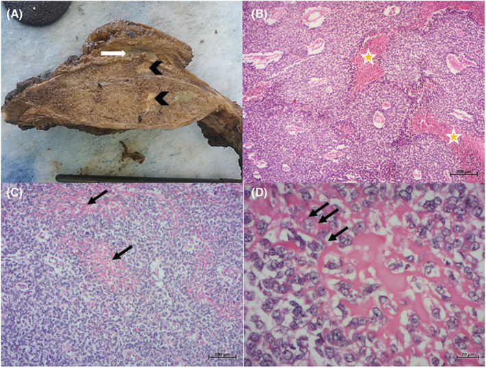
FIGURE 4.

Surgically resected specimen after neoadjuvant treatment: (A) at gross examination the sectioned specimen shows a large tan and hard tumor infiltrating the soft tissue (arrow) with few areas of yellowish necrosis (arrowhead); (B) microscopic examination shows a viable proliferation of uniform small round cells with rare foci of necrosis (asterisk) (H&Ex50); (C) closely packed tumor cells are interspaced with foci of osteoid production (arrows) (H&Ex100); (D) note the lace‐like osteoid deposited in between the uniform round cells (H&Ex400)
