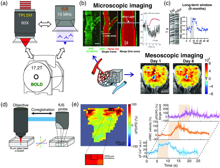
Fig. 2.
Previous use of PMP for small cranial windows in the olfactory bulb. (a) The same animal can be imaged with two-photon imaging, fUS and BOLD fMRI. (b) Two-photon (microscopic) and fUS (mesoscopic) imaging. Bottom left: schematic of the vascular network, which can be imaged at the microscopic (TPLSM, top) or mesoscopic level (fUS, right). Top: light per se dilates a pial artery labeled with Texas Red. Dilation follows calcium decrease in smooth muscle cells expressing GCaMP6f (scale bar: ) (modified from Rungta et al.18). Bottom: reproducibility of fUS responses () to odor (modified from Boido et al.11). (c) Red blood cell velocity response to odor in a capillary imaged (linescan acquisition) 9 months after the implantation of the window (unpublished data). (d) Coregistration of two-photon and fUS imaging systems: a given voxel is centered on a glass bead. (e) A specific voxel, centered on the glomerulus most sensitive to ethyl tiglate (left) shows an fUS response () that mirrors the increase of red blood cell velocity in the glomerulus capillary (modified from Aydin et al.19). Average of five responses for fUS and four responses for TPLSM imaging and red blood cell velocity.

