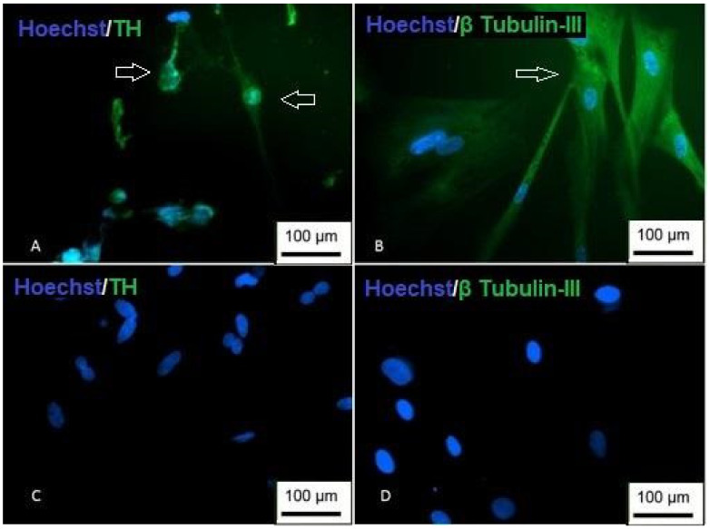
Figure 19.
Immunocytochemistry of dopaminergic differentiation. (A) Cells are showing nuclear staining (blue) and positive staining for TH (green); (B) cells with positive staining for β Tubulin-III; (C) undifferentiated TH control; (D) undifferentiated β Tubulin-III control (inversion optical microscopy—200 ×). Scale bar, 100 µm.

