Figure 1.
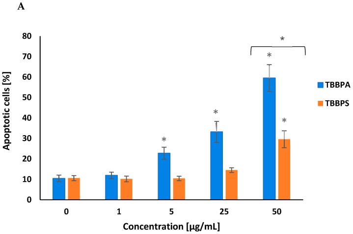
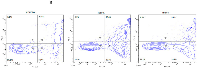
Apoptotic alterations in human PBMCs treated with TBBPA and TBBPS in the range from 1 to 50 µg/mL (n = 4) for 24 h (A). The cells were stained with Annexin-FITC and PI. Exemplary dot plots showing apoptotic alterations in human PBMCs unexposed (control) and exposed to TBBPA and TBBPS at 50 μg/mL for 24 h, Q3—live cells, Q2 + Q4—apoptotic cells (B). Statistically different from negative control at * p < 0.05. Statistical analysis was conducted using one-way ANOVA and a posteriori Tukey test.


