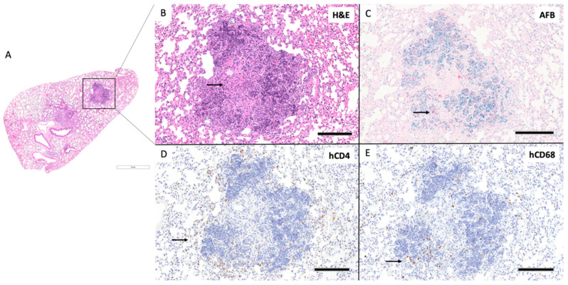
Figure 7.
HuDRAG-A2 mice show classically organized granuloma formation with human immune cell involvement at 4 weeks post-infected with H37Rv Mtb. (A) Whole lung section H&E (2×). (B) granuloma H&E (10×), arrow indicates foci of central caseating necrosis, (C) granuloma AFB (10×), arrows indicate Mtb bacilli stained red, (D) human CD4+ by IHC (10×), arrow indicates human CD4+ T cells stained brown, (E) human CD68+ by IHC (10×), arrow indicates human CD68+ macrophages stained brown; (scale bars for 2× = 1 mm; 10× = 200 µm).

