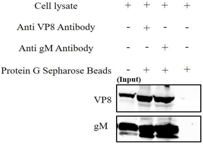
Figure 3.
VP8 and gM interact in WT BoHV-1-infected cells. Cell lysates from MDBK cells infected with WT BoHV-1 were incubated with VP8-specific or gM-specific antibodies, followed by Protein Sepharose G beads or with Protein G Sepharose without antibodies. After elution by SDS loading dye, the samples were subjected to SDS-polyacrylamide gel electrophoresis on an 8% gel and transferred to 0.45 µm nylon membranes. A fraction of the whole cell lysates was used as input control. VP8 and gM were detected with monoclonal anti-VP8 and rabbit anti-gM antibodies, followed by IRDye 680RD goat anti-mouse IgG and IRDye 800RD goat anti-rabbit IgG, respectively.

