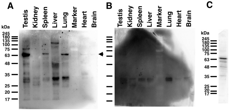
Figure 3.
Tissue distribution analysis of mouse HYAL4 protein in various mouse tissues. Expression levels of mouse HYAL4 protein in various tissues of a C57BL/6J mouse were examined by Western blotting using an anti-HYAL4 antibody (A-7), HRP-conjugate (A). HRP-conjugated normal mouse IgG was used instead of the primary antibody for the control experiments (B). HYAL4 protein expressed in L6 cells was detected by the HRP-conjugated anti-HYAL4 antibody A-7 as a positive control (C). The band detected at around 70 kDa indicated by an arrowhead on the right may be a glycosylated form of mouse HYAL4 protein (A). Bands detected at around 35 kDa may be nonspecific binding, as also detected in the control panel (B).

