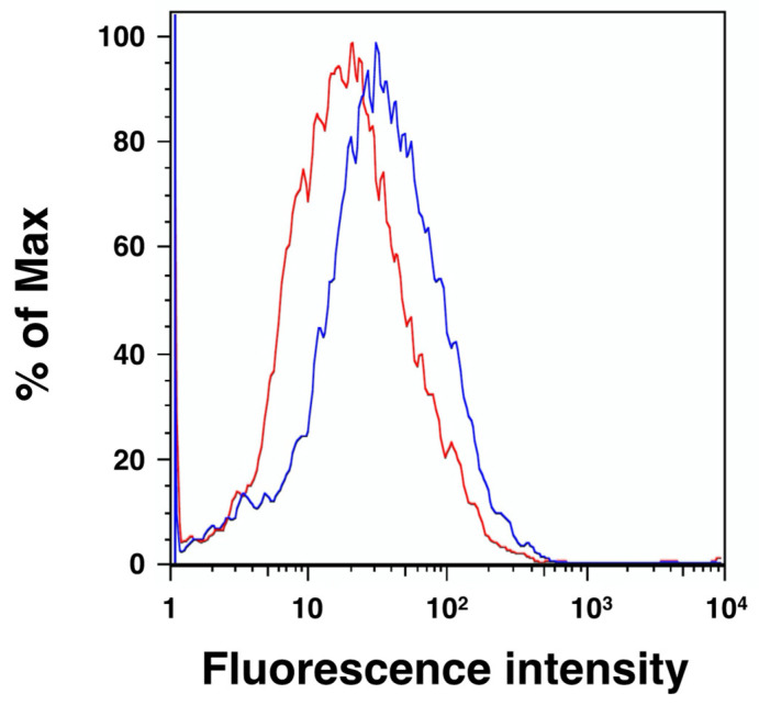
Figure 4.
Flow cytometric analysis of the HYAL4 protein on the surface of L6 cells. Representative line graphs of cellular staining are shown. Staining of L6 cells was performed using anti-HYAL4 antibody (clone A-7) (blue) or normal mouse IgG (red) under nonpermeabilized conditions. The binding of these antibodies to the epitopes on the cell surface was visualized by flow cytometry after incubating with Alexa Fluor 488-conjugated secondary antibody. Increased expression of HYAL4 on the cell surface was noted.

