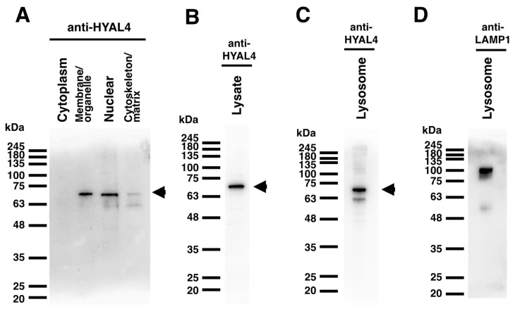
Figure 5.
Panel (A): Proteins in L6 cells were extracted and separated into cytosolic, organelle and membrane, nuclear, and cytoskeletal fractions using Subcellular Proteome Extraction Kit. An aliquot of each fraction was subjected to SDS-PAGE and transferred to a polyvinylidene difluoride (PVDF) membrane for blotting with the HYAL4 antibody. Panel (B): Whole cell lysate of L6 cells was subjected to Western blotting analysis with the HYAL4 antibody. Panels (C,D): Proteins in lysosomes of L6 cells were enriched and extracted by Lysosome Enrichment Kit. An aliquot was subjected to SDS-PAGE and transferred to a PVDF membrane for blotting with the HYAL4 antibody (C) and LAMP1 antibody (D). The band detected at around 70 kDa indicated by arrowheads on the right (A–C) may be a glycosylated form of mouse HYAL4 protein. When the normal IgG from mouse serum was used as the primary antibody, no bands were detected (results not shown).

