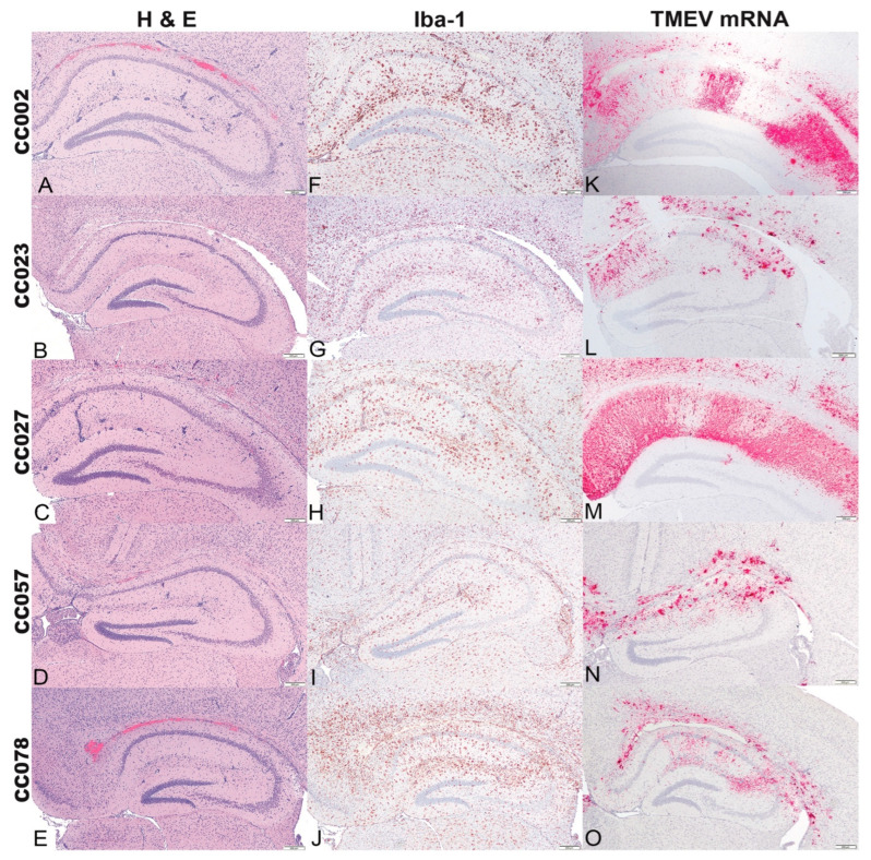
Figure 2.
Cross sections of the hippocampal formation at level B of CC mice infected with Theiler’s murine encephalomyelitis virus (TMEV) and euthanized at 4 dpi. (A–E) (Hematoxylin and eosin stain) Mild multifocal neuronal necrosis of CA1 and CA2 pyramidal layers with multifocal glial aggregates mostly centered around vessels (perivascular cuffing), predominantly in the stratum radiatum, stratum lacunosum moleculare. A linear area of hemorrhage in the alveus and hippocampal commissure was observed in A and E, interpreted as secondary to intracerebral TMEV inoculation. (F–J) (Iba-1 immunolabeling) All infected CC mice demonstrated increased numbers of Iba-1 positive microglia/macrophages in the hippocampal formation, most pronounced in the CC078 strain. (K–O) (TMEV RNA in situ hybridization) In all infected CC mice, mRNA expression was broadly distributed throughout the hippocampal formation. CC002 and CC027 strains had a radiating pattern of mRNA expression while CC023, CC057, and CC078 showed clustered patterns. Bar = 200 μm.

