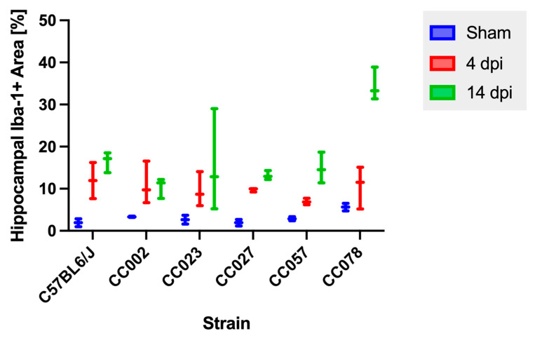
Figure 4.
Quantification of the Iba-1 immunopositive area within the hippocampal formation following intracranial inoculation with Theiler’s murine encephalomyelitis virus. No mice showed a significant change in the Iba-1+ area between 4 dpi and 14 dpi. p values were determined using the Wilcoxon rank sum tests.

