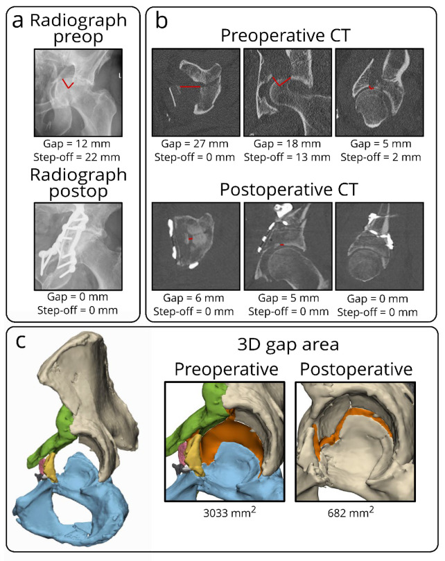
Figure 3.
Case example of a both-column fracture in a 63-year-old male, showing the discrepancy in measuring initial and residual fracture displacement for acetabular fractures on different imaging modalities, including radiographs, CT scans, and 3D models. On radiographs (a), it is difficult to measure gaps and step-offs, especially on the postoperative radiograph, because the implant is partially obscuring the acetabulum. On the single CT slices (b), multiple gaps and step-offs (red lines) can be measured on different CT slices in several planes, indicating the subjective elements of these measurements. The 3D model (c) demonstrates the 3D gap area (in orange) representing the three-dimensional surface between all fracture fragments. This should be considered a single quantitative measure of the initial or residual fracture displacement in the entire acetabulum.

