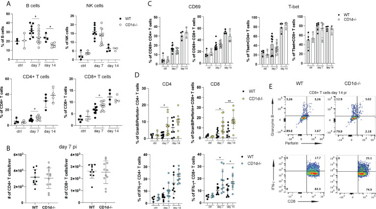
Figure 5.
Hepatic lymphocyte subsets in CD1d-/- mice during acute infection. A flow cytometric analysis of hepatic lymphocytes in WT and CD1d-/- mice (n=4-8 per time point) was performed at day 7 and 14 pi and in uninfected controls. (A) Frequencies of B cells, NK cells, CD4+ and CD8+ T cells. (B) Total numbers of hepatic CD4+ and CD8+ T cells at day 7pi. (C) CD69 and T-bet expression of CD4+ and CD8+ T cells. (D) Granzyme B and perforin (upper graphs) and IFN-γ (lower graphs) expression of T cell subsets after unspecific stimulation with PMA/Ionomycin. (E) Representative FACS plots of granzyme B/perforin and IFN-γ expression in CD8+ T cells at day 14 pi. Unpaired t test: *p< 0.5, **p<0.01.

