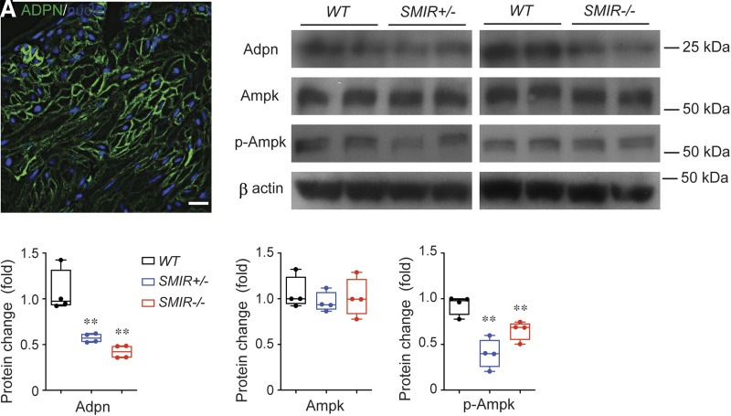
Figure 7.
Inhibition of adiponectin signaling in SMIR+/− and SMIR−/− mice bladders. A: Immunostaining and imaging of adiponectin (green) in BSM cell membrane. Nuclei are stained with DAPI (blue). B: Western blot of Adpn, Ampk, and p-Ampk proteins from wild-type (WT), SMIR+/−, and SMIR−/− mice bladders (n = 4). β-Actin is used as loading control for normalization, and quantitated data are shown in C–E. Data are shown as boxes and whiskers. The centerline is the median of the data set, the box represents 75% of the data, and bars indicate whiskers from minimum to maximum. Data were analyzed with use of Student t test. *P < 0.05 and **P < 0.01.

