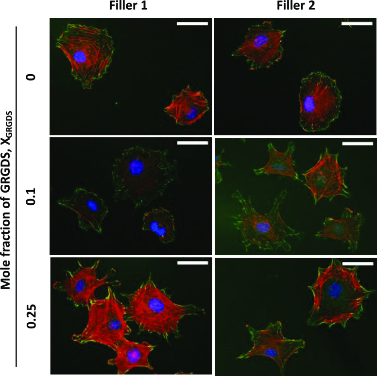
Figure 3.
Double immunofluorescence labeling. Representative images illustrate the effect of GRGDS 3 density and filler amphiphiles on the morphology of MC3T3-E1 cells adhered to rSAMs anchored on MBA-SAMs. The labeling used to visualize the cells are nucleus (DAPI: blue), focal adhesions (phospho-paxillin: green), and actin filaments (phalloidin: red). The images were recorded 5 h after seeding. Scale bars = 50 μm.

