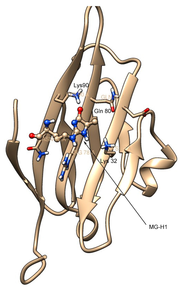
Figure 6.
Solution NMR structure of the RAGE-MG-H1 complex (shown here is the extracellular V-domain of the RAGE bound to MG-H1); the imidazolone moiety of MG-H1 is surrounded by the positively charged amino acid residues, including Lys 90, Gln 80, and Lys 32; the structure was created using UCSF Chimera software; PDB ID: 2MOV (MG-H1 is shown as a ball-and-stick model; red = oxygen, blue = nitrogen).

