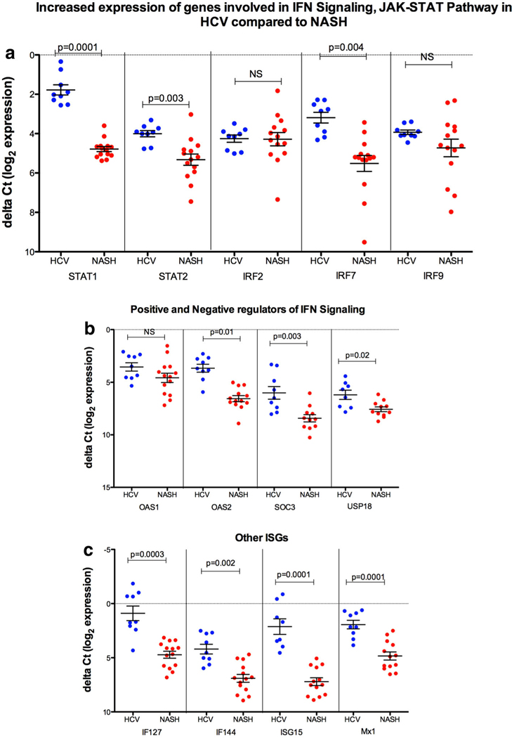
Fig. 2.
a, b IFN signaling and JAK-STAT pathway such as STAT1, STAT2 and IRF7 SOCS3, OAS2, and USP18 (negative regulator), were highly expressed in HCV GT-3 versus NASH. c Other important ISGs such as IFI27, IFI44, Mx1 and ISG15 were significantly increased in HCV GT-3 compared to NASH. Values are mean ± SE of the delta Ct values. p value was calculated by the unpaired t test with Welch’s correction using GraphPad Prism 6.0 software. p values less than 0.05 were considered significant

