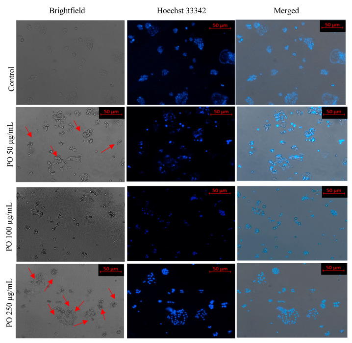
Figure 7.
Nuclear morphology of MCF-7 cells after DAPI staining. The fluorescence microscopy images (at 10×) of cells treated with different concentrations of EEPOs show condensed nuclei, high fluorescence and cell shrinkage as compared to the negative control, which does not exhibit such nuclear changes. Red arrowhead: Cell apoptosis in treatment group.

