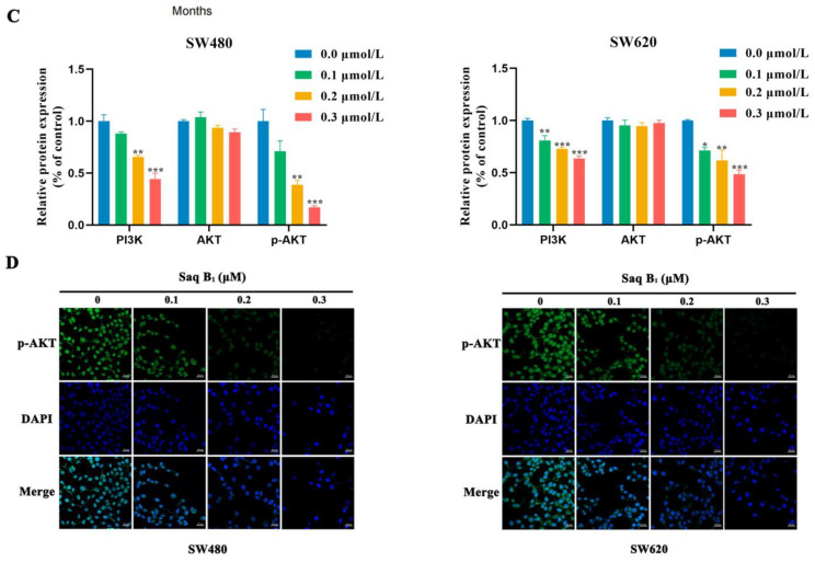
Figure 3.
Effect of Saq B1 against the PI3K/AKT pathway in SW480 and SW620 cells. (A) The overall survival rate of patients with high or low PIK3CA expression was assessed via Kaplan–Meier survival analysis. (B,C) Western blot analysis was used to investigate the effects of Saq B1 on PI3K, AKT, and p-AKT proteins. (D) The expression of p-AKT was observed by immunofluorescence staining. Representative images of each condition are shown; p-AKT (green) and DAPI (blue). Scale bars = 20 µm. The level of intracellular p-AKT decreased, which was consistent with Western blot results. * p < 0.05, ** p < 0.01, *** p < 0.001.


