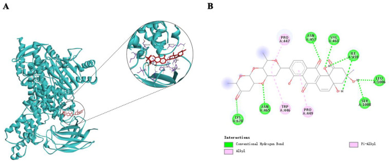
Figure 4.
Theoretical binding mode of Saq B1 to PI3Kα protein. (A) AutoDockTools software was used to obtain the optimal docking model of Saq B1 to the PI3Kα protein, and the image obtained was a 3D panorama of the optimal binding mode. (The chemical structure of Saq B1 is represented by a red rod-shaped structure, the blue band represents the spatial structure of the PI3Kα protein, and the purple rod-shaped structure represents the amino acid residue bound to Saq B1). The lowest binding energy configuration is −8.34 kcal/mol. (B) The simplified two-dimensional interaction model is presented, in which some carbonyl and hydroxyl groups of Saq B1 are hydrogen-bonded to amino acid residues, while the D-olivose and benzene ring are bonded to amino acid residues Pro447, Trp446, and Pro449 in the form of alkyl.

