Figure 5.
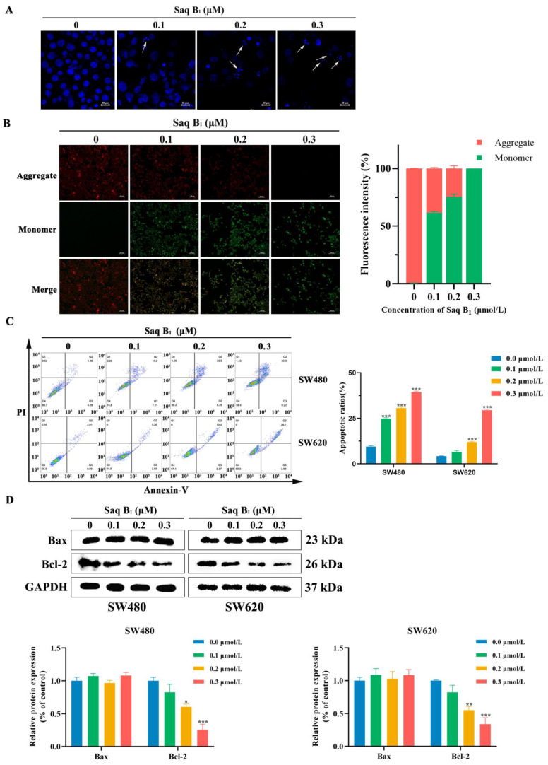
Saq B1 induces apoptosis in CRC cells. (A) DAPI staining was used to study the effect of Saq B1 on apoptosis in SW480 cells. The arrows show nuclei that are condensed and fragmented within the cell. Scale bars = 20 µm. (B) Changes in MMP were detected using the JC-1 probe after SW480 cells were treated with different concentrations of Saq B1. Representative images are shown. Scale bars = 100 µm. The fluorescence intensity of JC-1 was analyzed using Image J software, and the statistical graph was drawn. (C) Apoptosis in SW480 and SW620 cells treated with and without Saq B1 was determined by flow cytometry using Annexin V-FITC/PI staining. (D) Western blot was used to analyze the expression levels of Bax and Bcl-2. * p < 0.05, ** p < 0.01, *** p < 0.001.

