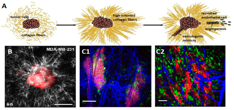
Figure 3.
Tumor cells organize the disordered collagen fibers into orientation, further providing a scaffold to recruit endothelial cells (green cell in illustration) for angiogenesis or inducing vasculogenic mimicry constituted by tumor cells (red cell in illustration) without endothelial cells presenting. (A) The cartoon diagram displays the process of angiogenesis. (B) A confocal image of a representative tissue comprised of breast cancer cell MDA-MB-231. The collagen fibers around the tissue are aligned, obviously. Reprinted/adapted with permission from Ref. [76]. (C1,C2) The SHG images show the vasculature (blue) formed neighbor tumor within the micro-environment of high dense collagen matrix. Reprinted/adapted with permission from Ref. [74].

