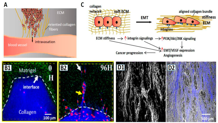
Figure 4.
Collagens promote cancer invasion. (A) The schematic representation of oriented collagen fibers providing a “highway” to guide cancer cells in realizing the intravasation process. (B1,B2) The fluorescent images show the stiffness Matrigel (green) region is invaded by cancer cells (red) in the guidance of the oriented collagen fibers (blue) [59]. (C) The schematic representation of stiffness ECM inducing EMT accompanied with high adhesion of tumor cells. (D1,D2) Tumor cells (gray color in (D2)) invade along with the direction of collagen fibers. (D1) The SHG signal of collagen fibers in the pathological section. (D2) The merge of the light field and SHG signal of the pathological section [59].

