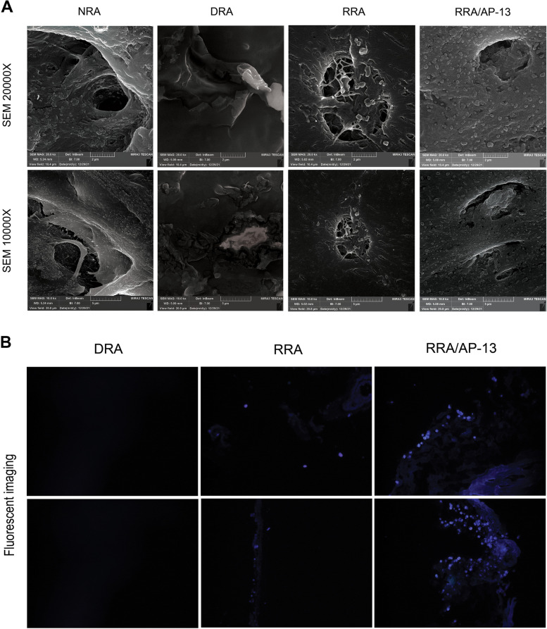
Fig. 7.
FESEM analysis of the luminal side of NRA, DRA, RRA, and RRA/AP-13 demonstrates uniform repopulation of acellular scaffold after the recellularization process and higher cell attachment to RRA/AP-13 tissue compared to RRA (A). The presence of a higher number of cells stained by Hoechst 33342 stain in RRA/AP-13 tissue demonstrates increased efficacy of cell attachment to RRA/AP-13 scaffold compared to the RRA tissue. The first and second rows represent different sides of tissue (B). NRA: Native Rat Aorta, DRA: Decellularized Rat Aorta, RRA: Recellularized Rat Aorta, RRA/AP-13: Recellularized Rat Aorta Conjugated with Apelin-13

