Fig. 1.

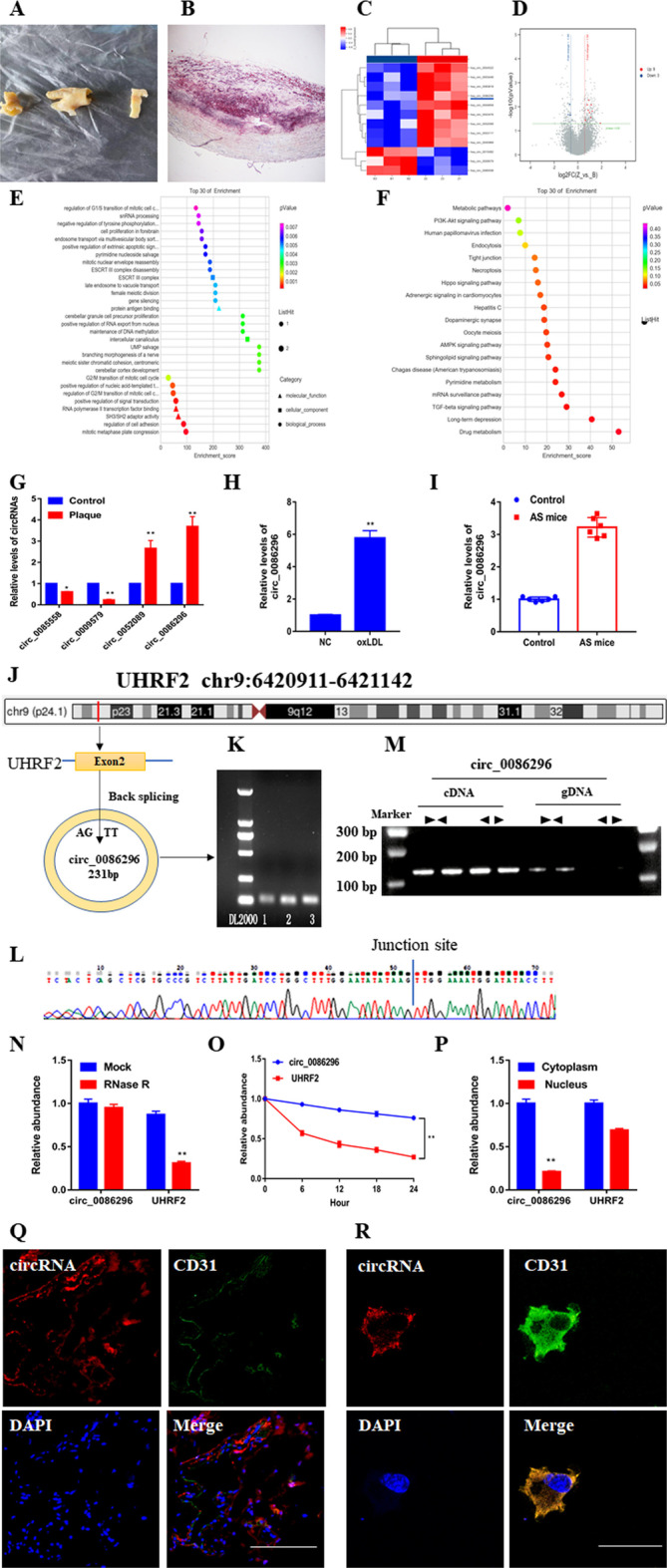
Validation and characterization of circ_0086296. A Morphology of human carotid atherosclerotic plaque tissues. B Human plaque tissue sections were stained with Oil Red O. C Cluster heatmap showing the abnormally expressed circRNAs from the microarray data. D Volcano map showing the abnormally expressed circRNAs. E Gene Ontology analysis of the abnormally expressed circRNAs. F Abnormally expressed circRNAs were identified by Kyoto Encyclopedia of Genes and Genomes pathway analysis. G The expression of abnormally expressed circRNAs in plaque samples. H, I Levels of circ_0086296 in HUVECs treated by oxidized low-density lipoprotein (ox-LDL) (100 μg/mL) for 24 h (H) and in the aorta of atherosclerotic mice (I) were determined via quantitative real-time polymerase chain reaction (qRT-PCR). J Graphic showing UHRF2 circularization to form circ_0086296. K The results of circ_0086296 PCR using agarose gel electrophoresis. L The back-splice junction sequences of circ_0086296 were found using Sanger sequencing. M–P circ_0086296 expression in HUVECs (M); in HUVECs after RNase R treatment (N); levels of circ_0086296 and UHRF2 after actinomycin D treatment (O); and levels of circ_0086296 and UHRF2 mRNA in the cytoplasm and nucleus of HUVECs (P) were determined via qRT-PCR. Q, R The localization of circ_0086296 in plaque tissues (scale bar, 50 μm) (Q) and localization of circ_0086296 in HUVECs (scale bar, 10 μm) (R) were verified via fluorescence in situ hybridization (FISH). Nuclei were stained with DAPI (blue), and circ_0086296 probes were labeled with Cy3 (red). *p < 0.05; **p < 0.01 versus the relative control group
