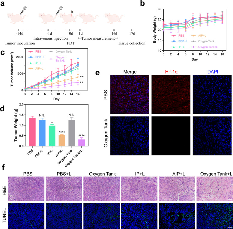
Fig. 5.
In vivo anti-tumor effect of the Oxygen Tank. a Schematic illustration of the PDT treatment (1 W cm − 2, 30s) after tail vein injection (200 uL, 100 μg/mL IR780, n = 6). b and c Body weight and tumor volume curves. d Weight of tumors. e Hif-1α staining tumor sections. The scale bar is 50 μm. f Photographs of the H&E and TUNEL staining of the AGS-bearing mice in different treatments (PBS, PBS with laser, Oxygen Tank, IP NPs with laser, AIP NPs with laser, and Oxygen Tank with laser). The scale bars are 100 μm. Data are shown as mean ± SD. *p < 0.05, **p < 0.01, ***p < 0.001, ****p < 0.0001, while N.S. means Not Significant

